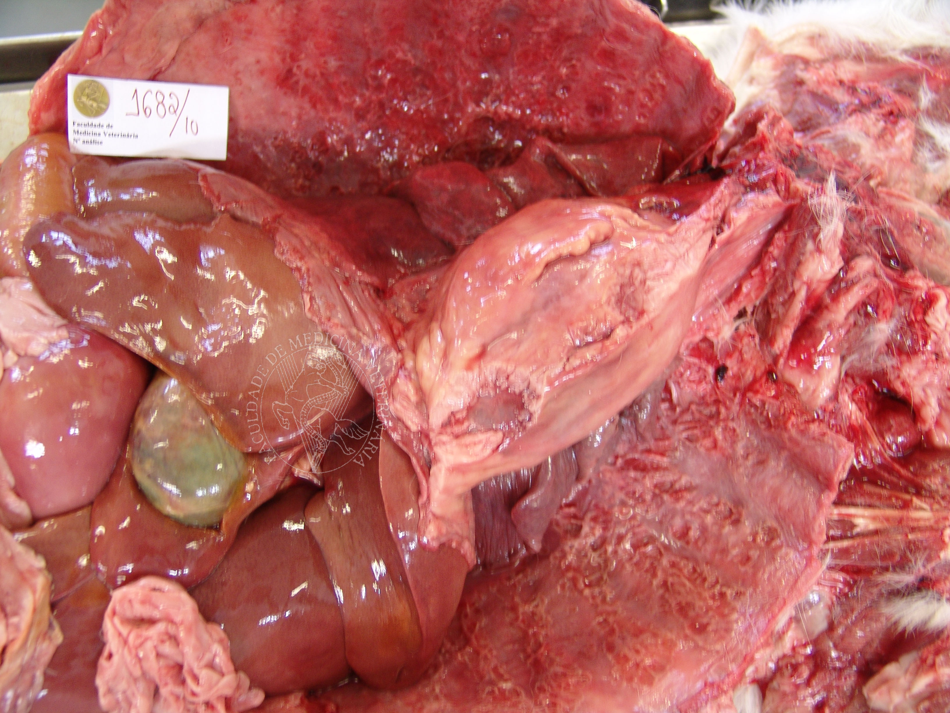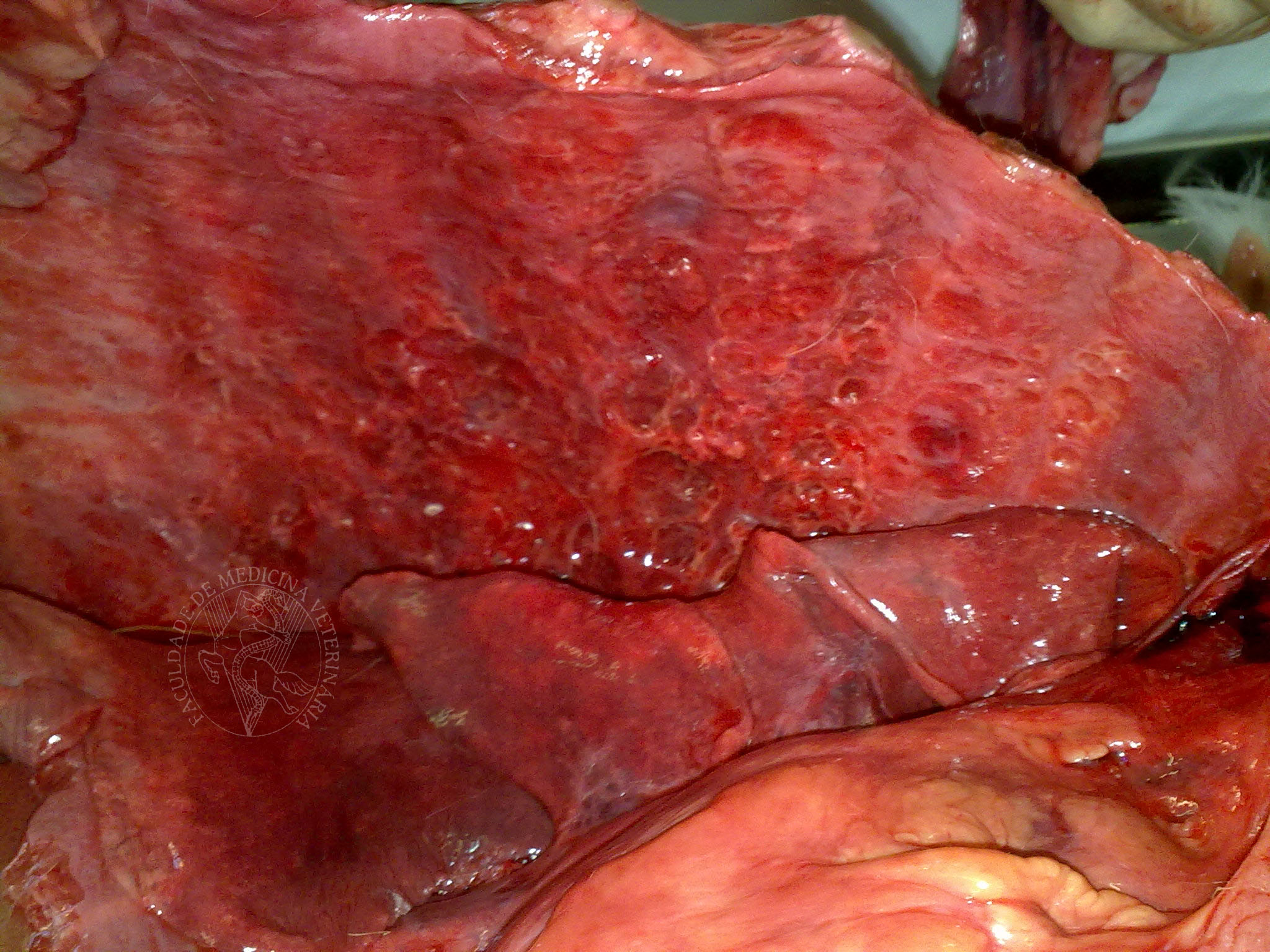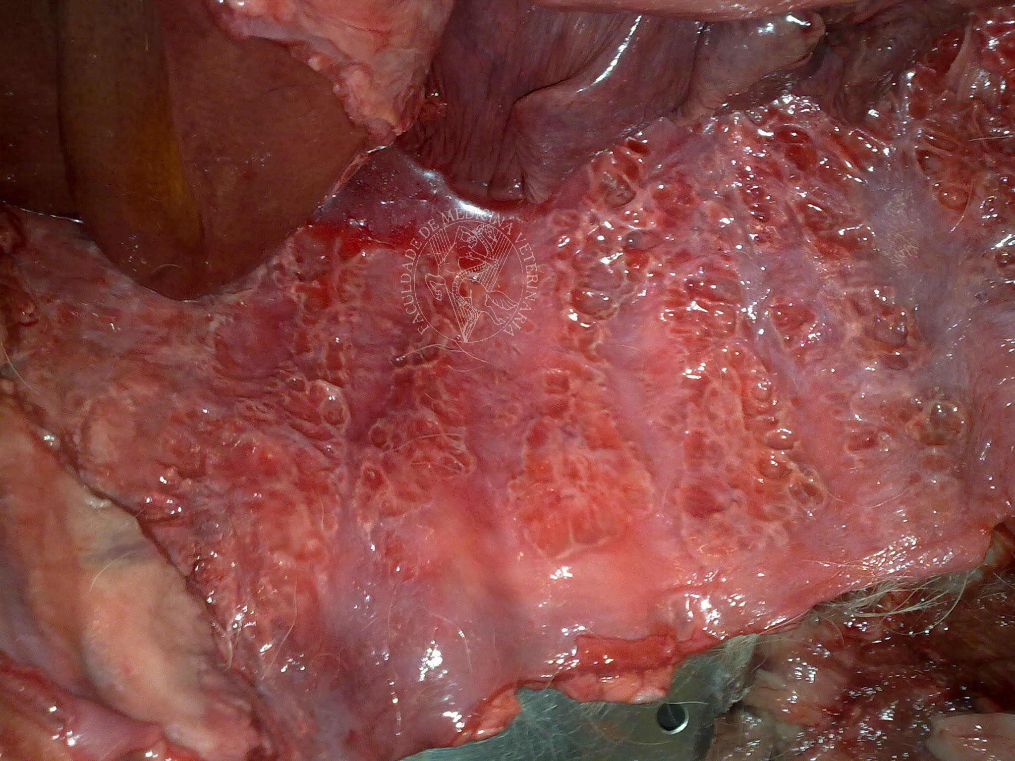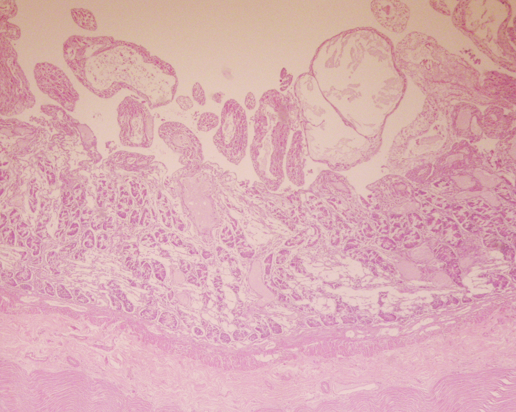

Pleural mesothelioma in a 12-year-old Golden Retriever. This animal’s thoracic cavity was full with around 1L of blood-stained fluid. Note the slightly bulging formations of hard consistency and pale contours, which completely line the thoracic wall, with a “geographic map” type profile. Both lungs are collapsed, with an irregular surface and adherences to the diaphragm. Histopathologically, the lesions in the parietal pleura corresponded to neoplastic proliferation of mesothelial cells, formed by finger-like structures triggering exuberant osseous and cartilaginous metaplasia of the supporting conjunctive tissue.
 |
 |
 |
 |
 |
 |
Portuguese
English
 |
 |
 |
Loading
Copyright © 2001 by MC Peleteiro. M Pinho & JS Orvalho
design by R Noiva