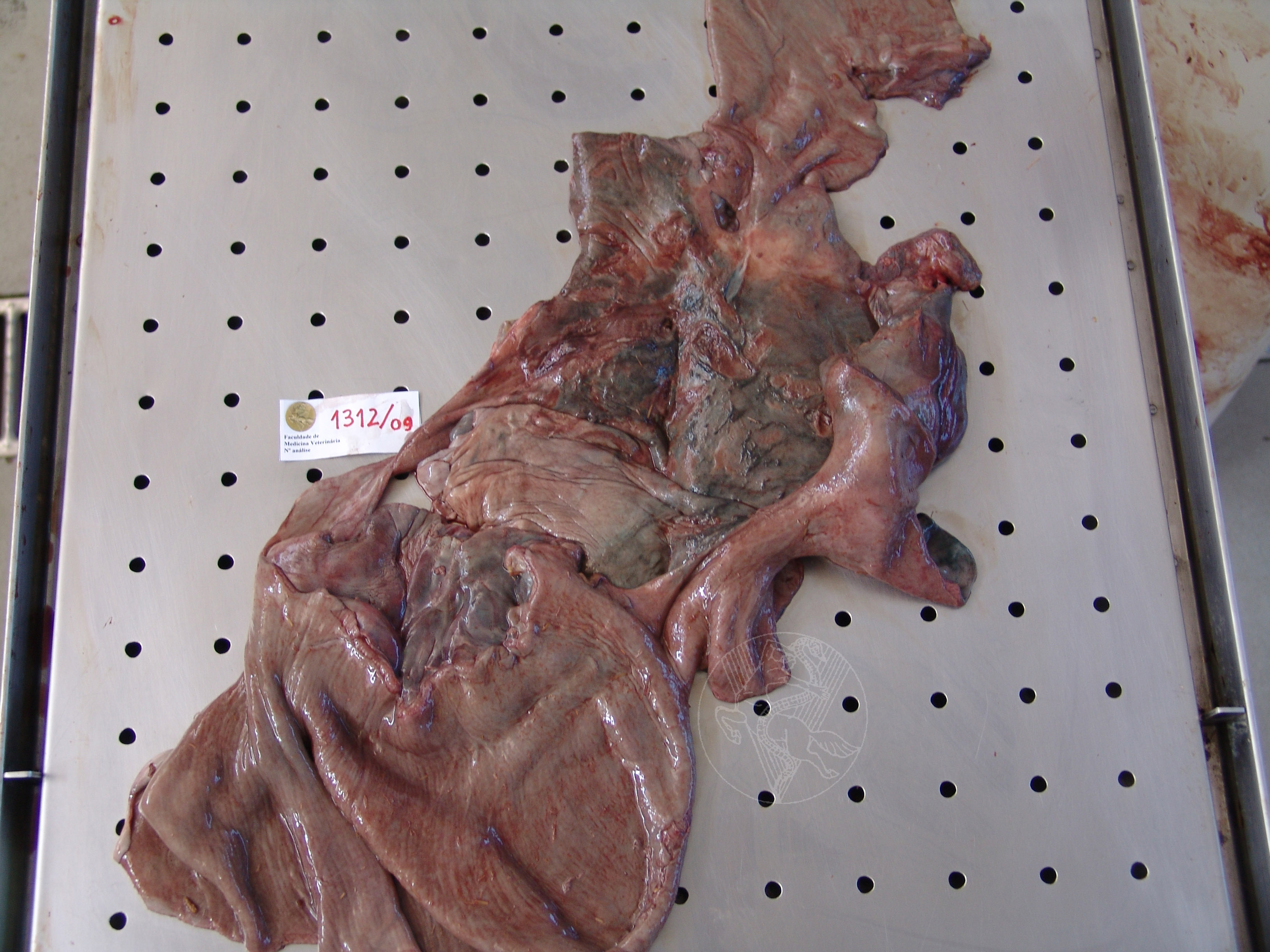

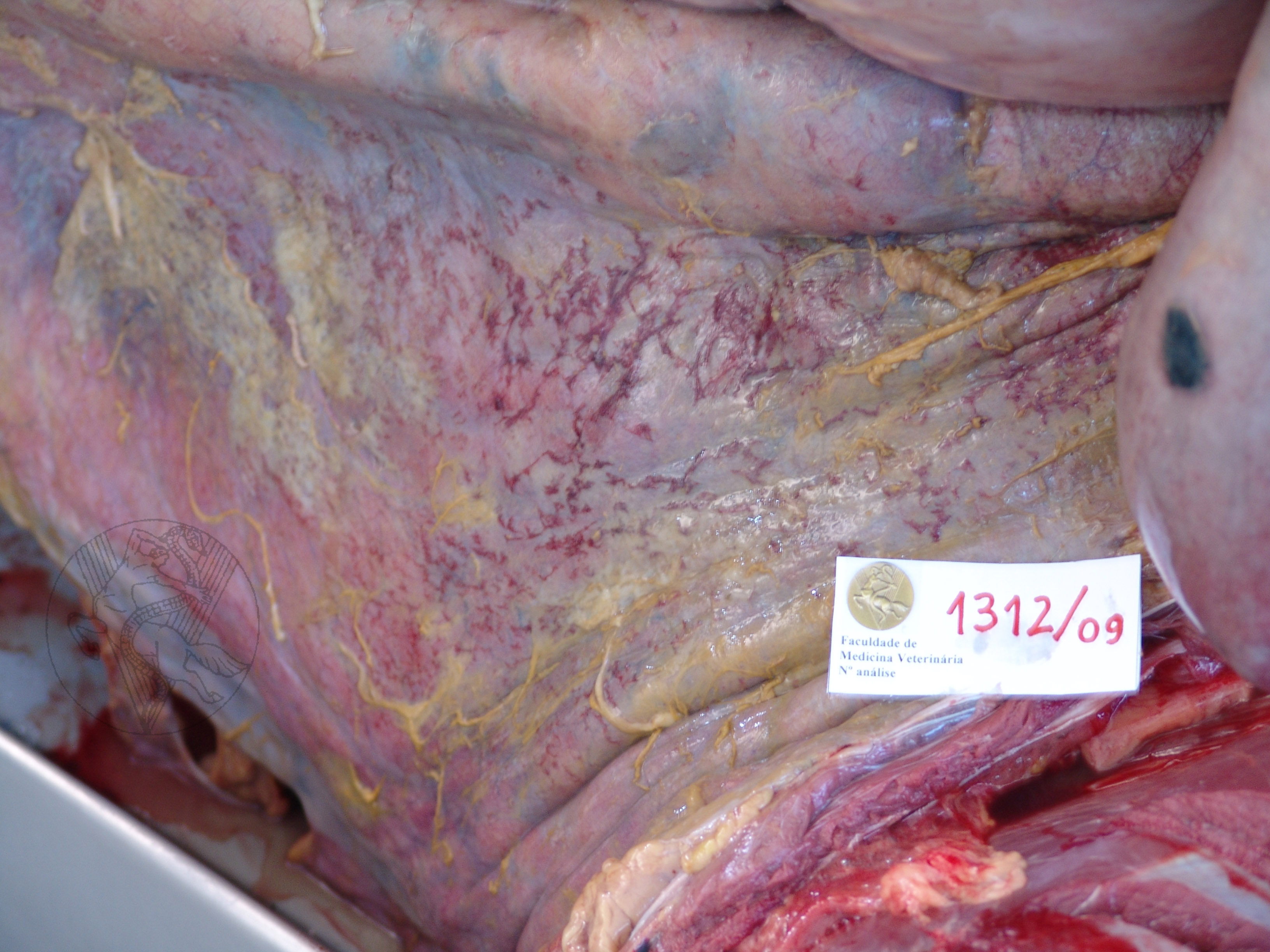
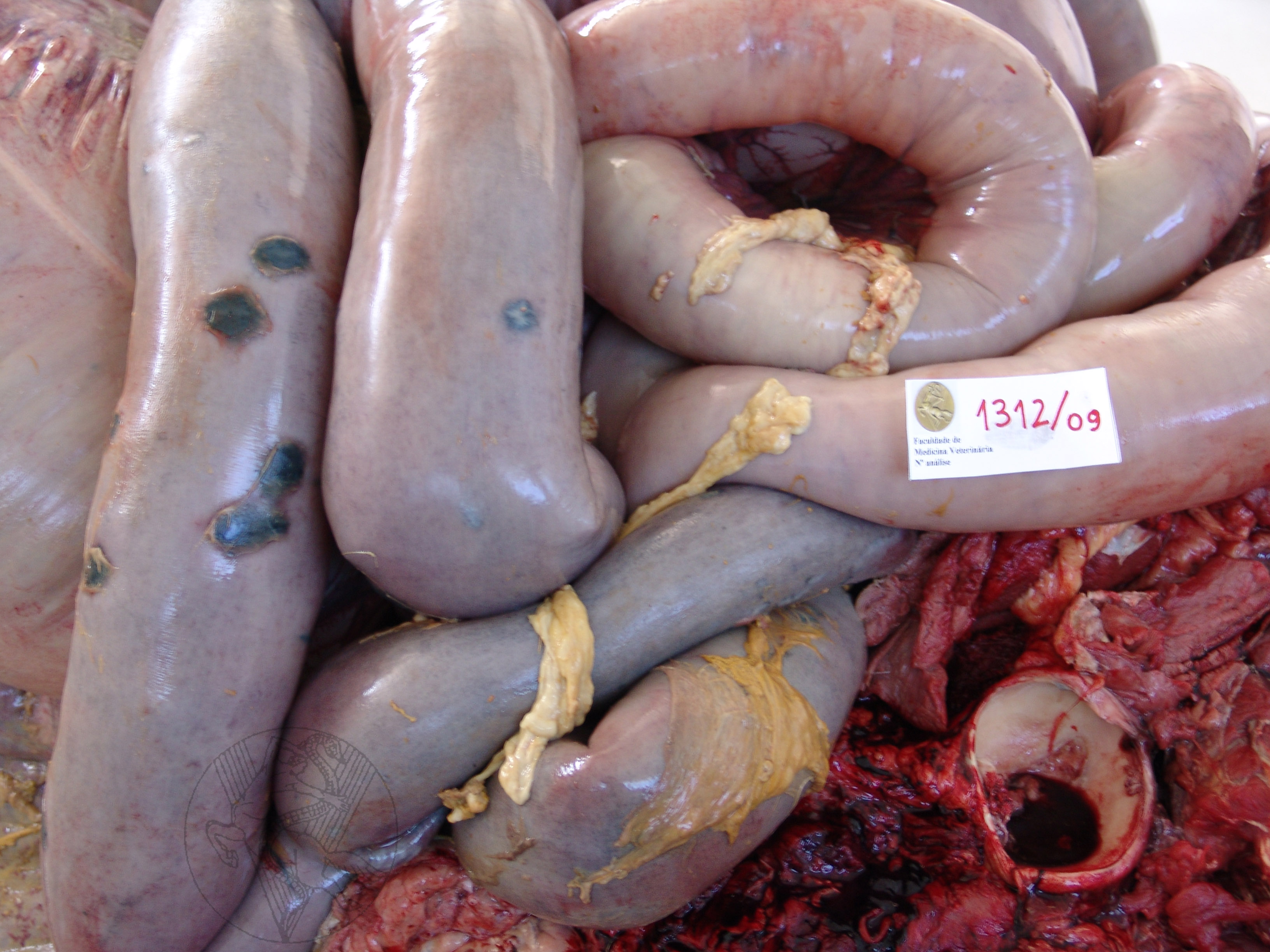
Fibrinous peritonitis in a 12-year-old cross-breed Lusitan. Congestion and fibrin filaments can be seen. On the right, are foci of chronic peritonitis on the colonic wall, corresponding to areas of serosal thickening darkened by impregnation with haematic pigment. Below, note the mass that was located at the entrance of the pelvic cavity. Its dissection revealed it to result from the formation of a large, multilocular cavity of unknown nature that was apparently attached to the intestinal lumen. Histological analysis proved this case to be one of intestinal rupture that must have taken place several months prior to death. The omentum was involved in limiting the dispersion of intestinal contents, contributing to the slow evolution of an initially focal peritonitis.
 |
Portuguese
English
 |
 |
 |
Loading
Copyright © 2001 by MC Peleteiro. M Pinho & JS Orvalho
design by R Noiva