

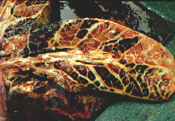
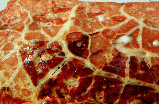
Contagious peripneumonia in a cow. The cut surface of the lung exhibits the marked heterogeneity of the lesions that grants the lung a distinctive marble-like appearance. Note the very evident oedema of the interlobular septa. The image on the right shows the septal distension in greater detail, where thrombosed lymphatic vessels (arrow) as well as foci of perivascular organization (circles) can be seen. The first arise as elongated structures of clear content, sometimes occupying more than half of the total diameter of the septum in which they are inserted. The latter, appear as small red dots corresponding to blood vessels surrounded by a halo of lardaceous tissue.
 |
 |
 |
 |
 |
 |
Portuguese
English
 |
 |
 |
Loading
Copyright © 2001 by MC Peleteiro. M Pinho & JS Orvalho
design by R Noiva