
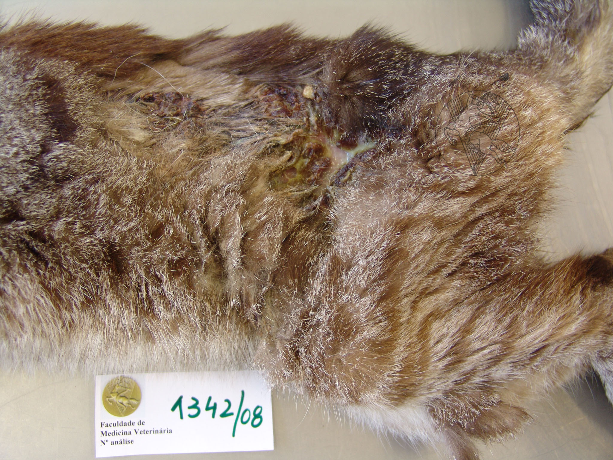
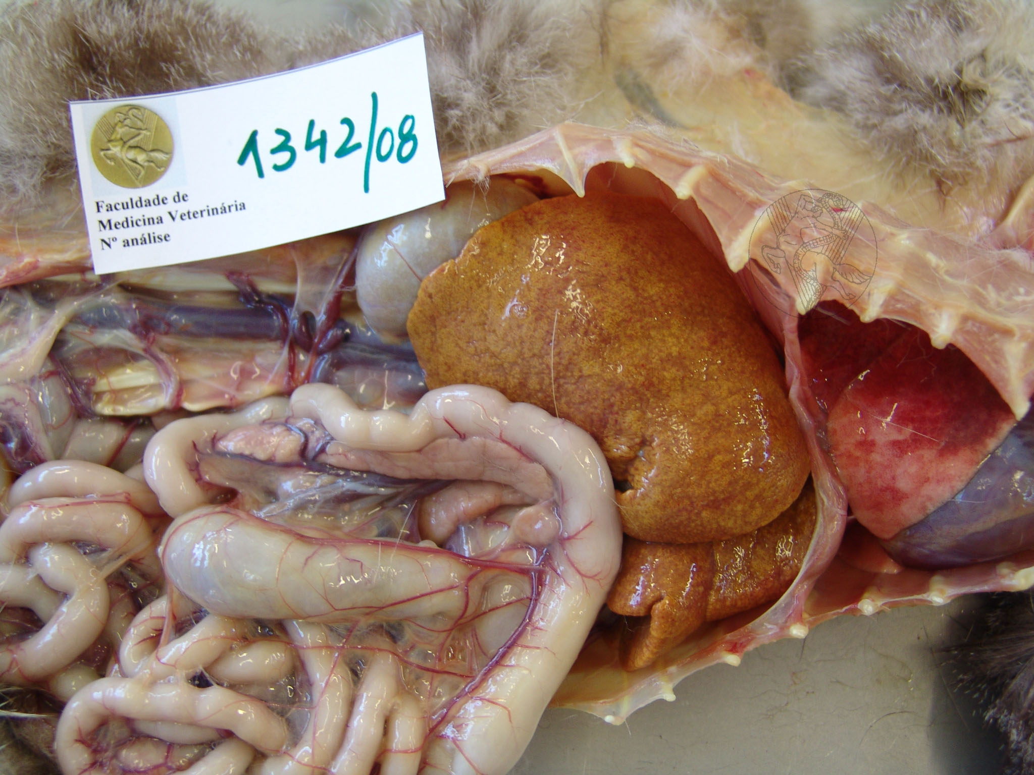
Skin fragility syndrome in a cat. The dorsal and lumbar regions exhibit extensive crusted lesions with underlying purulent material. This animal’s liver (on the right) presented with an intense yellow colouration, corresponding to a severe degenerative lesion.
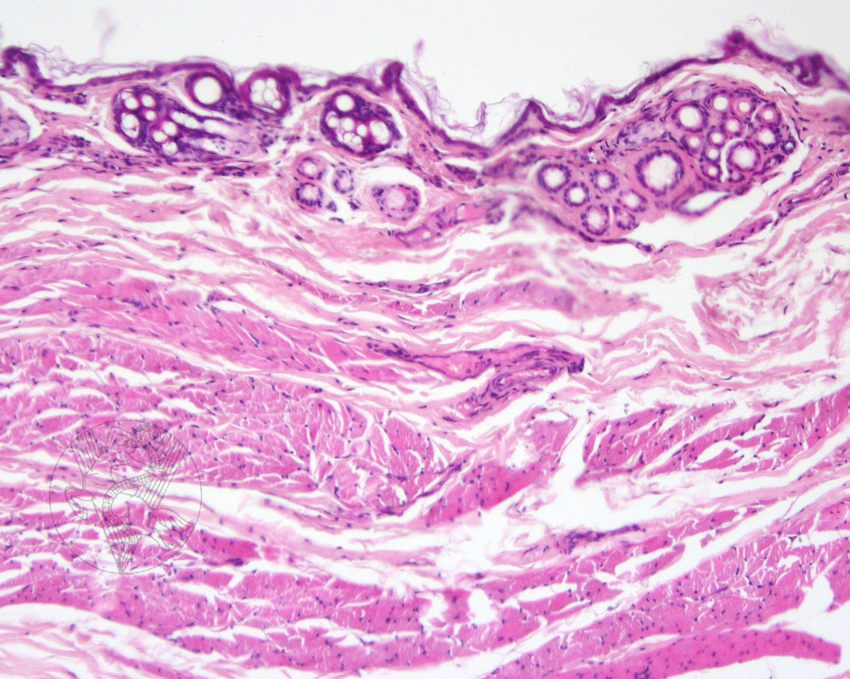
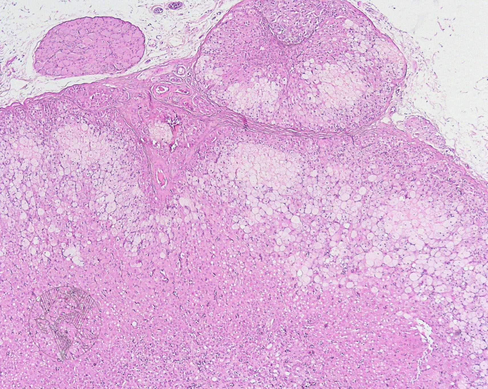
Below, on the left, a microscopy image of the skin is displayed. There is marked atrophy of the dermis, with hair follicles appearing close together in a very small area. This cat was also diagnosed with apparently secondary hyperadrenocorticism, although no hypophyseal tumour has been detected. A microscopy image of an adrenal gland can be seen on the right, showing marked hypertrophy of the cortical cells, which cytoplasm appears finely granulous. Note the small cortical nodule that invades the extracapsular space, corresponding to cortical hyperplasia. The extracapsular nodule that can be seen above and to the left is a ganglion.
 |
 |
Portuguese
English
 |
 |
 |
Loading
Copyright © 2001 by MC Peleteiro. M Pinho & JS Orvalho
design by R Noiva