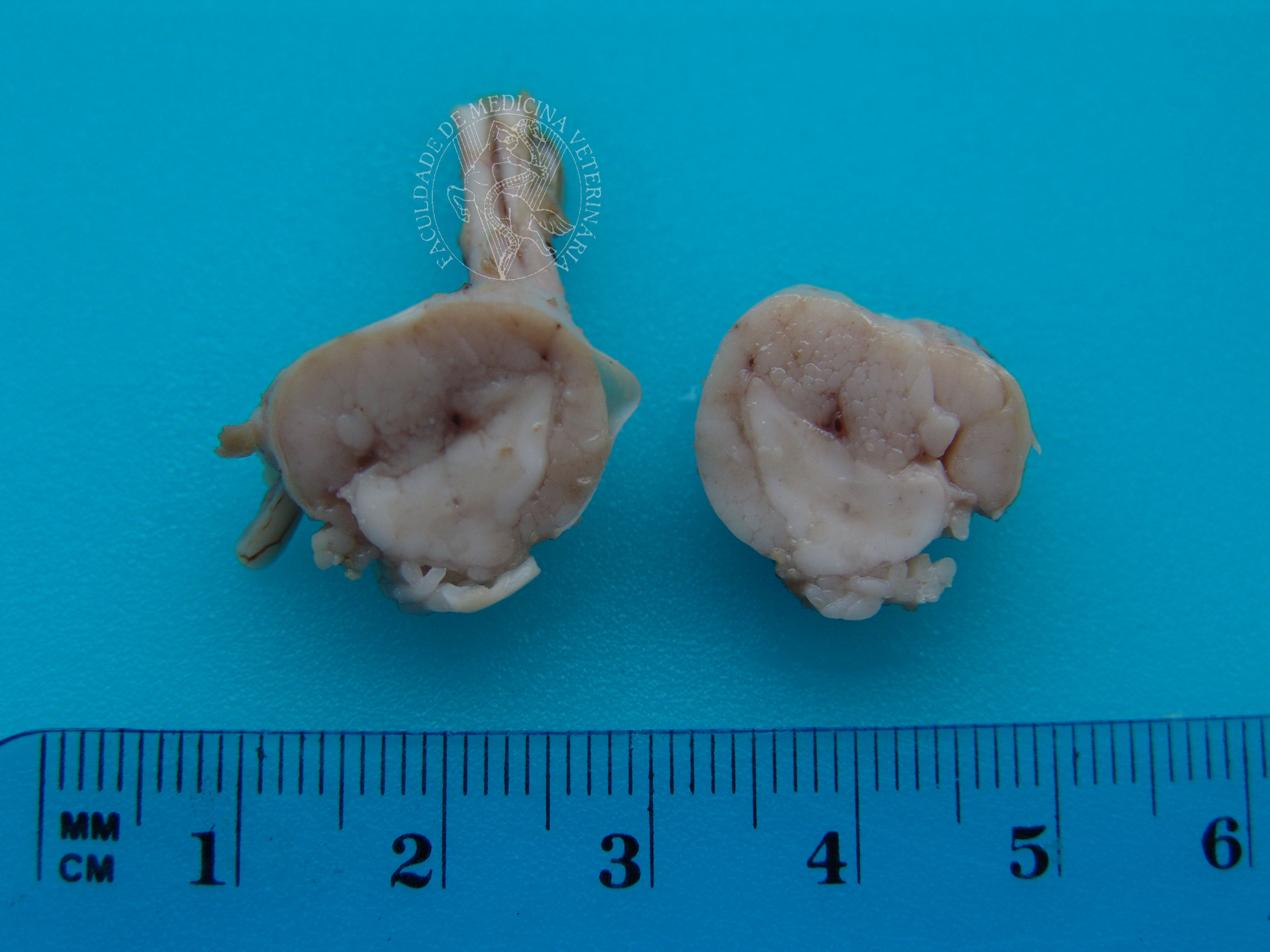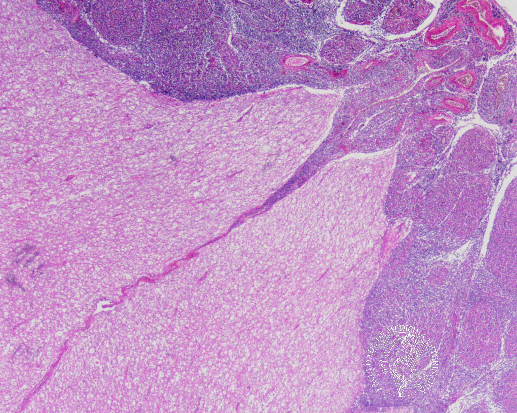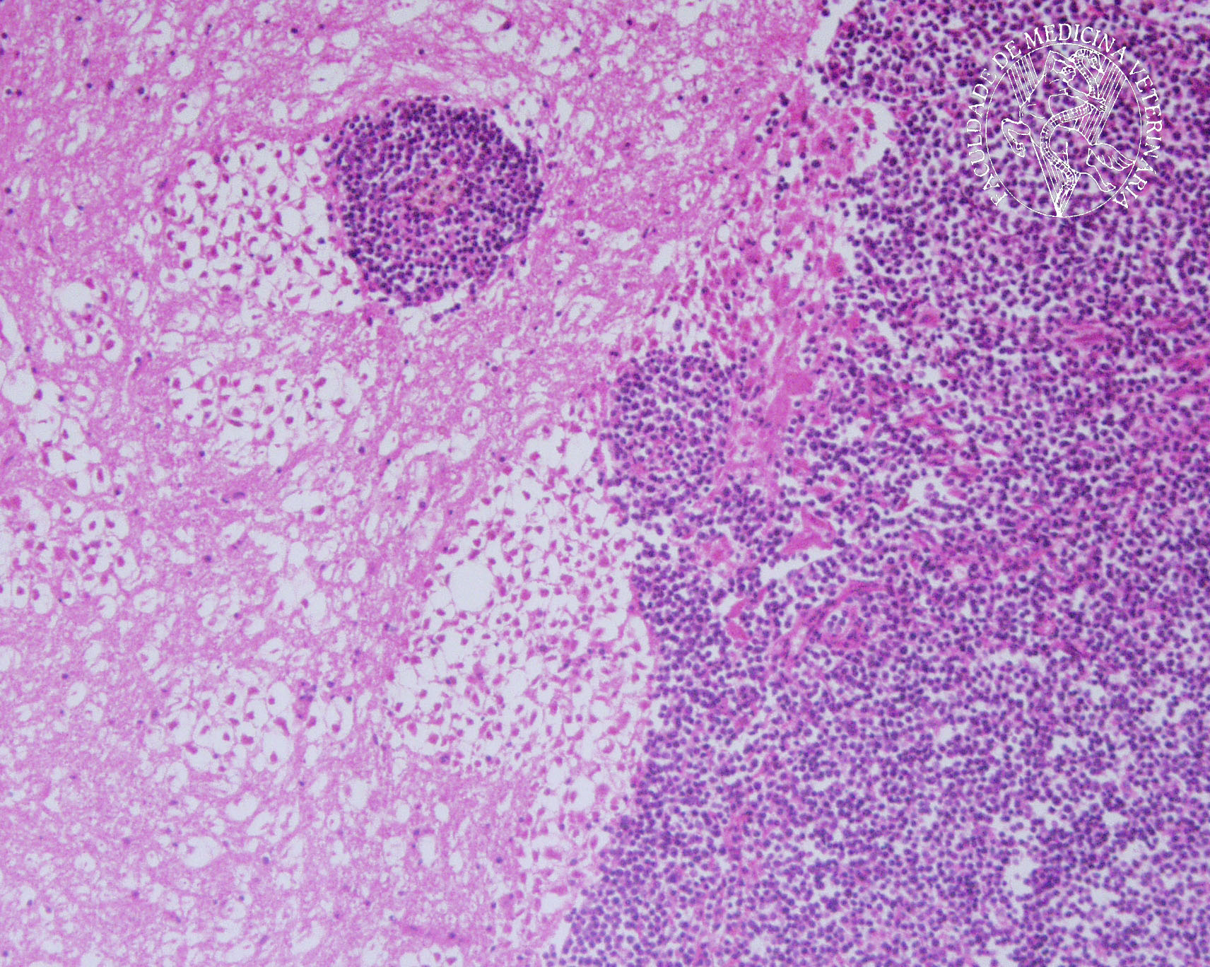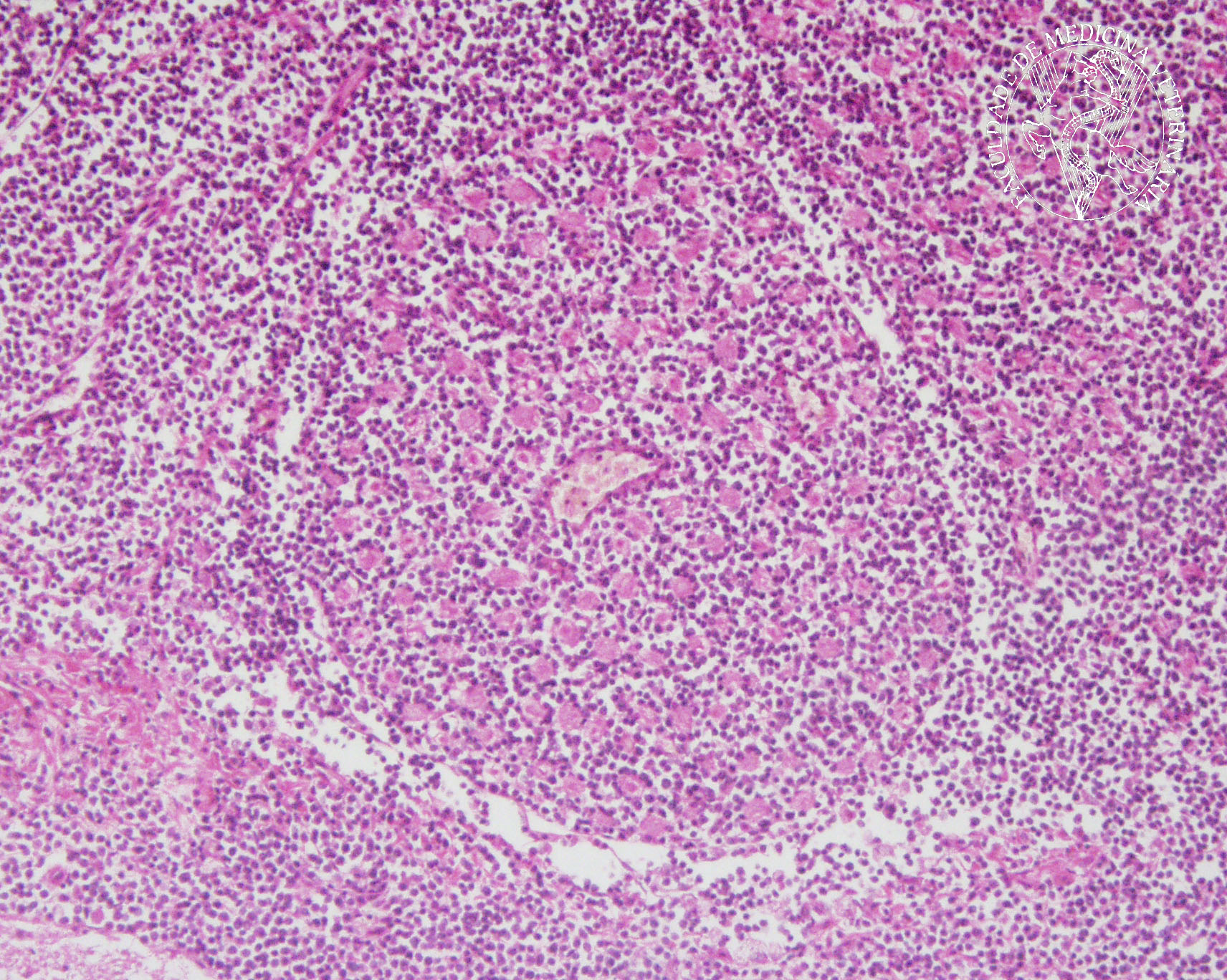

Solitary subdural lymphoma in a 13-year-old Serra da Estrela crossbreed. Upon opening the spinal canal, the spinal cord appeared thickened over the L4 and L5 vertebrae. A sagittal cut exposed the severe compression and deformation of the spinal cord (whitish in the image) by neoplastic lardaceous tissue that infiltrated and surrounded the chord and emergent rachidian nerves. Histology and immunohistochemistry revealed the neoplastic cells to be T-lymphocytes.
 |
 |
 |
Portuguese
English
 |
 |
 |
Loading
Copyright © 2001 by MC Peleteiro. M Pinho & JS Orvalho
design by R Noiva