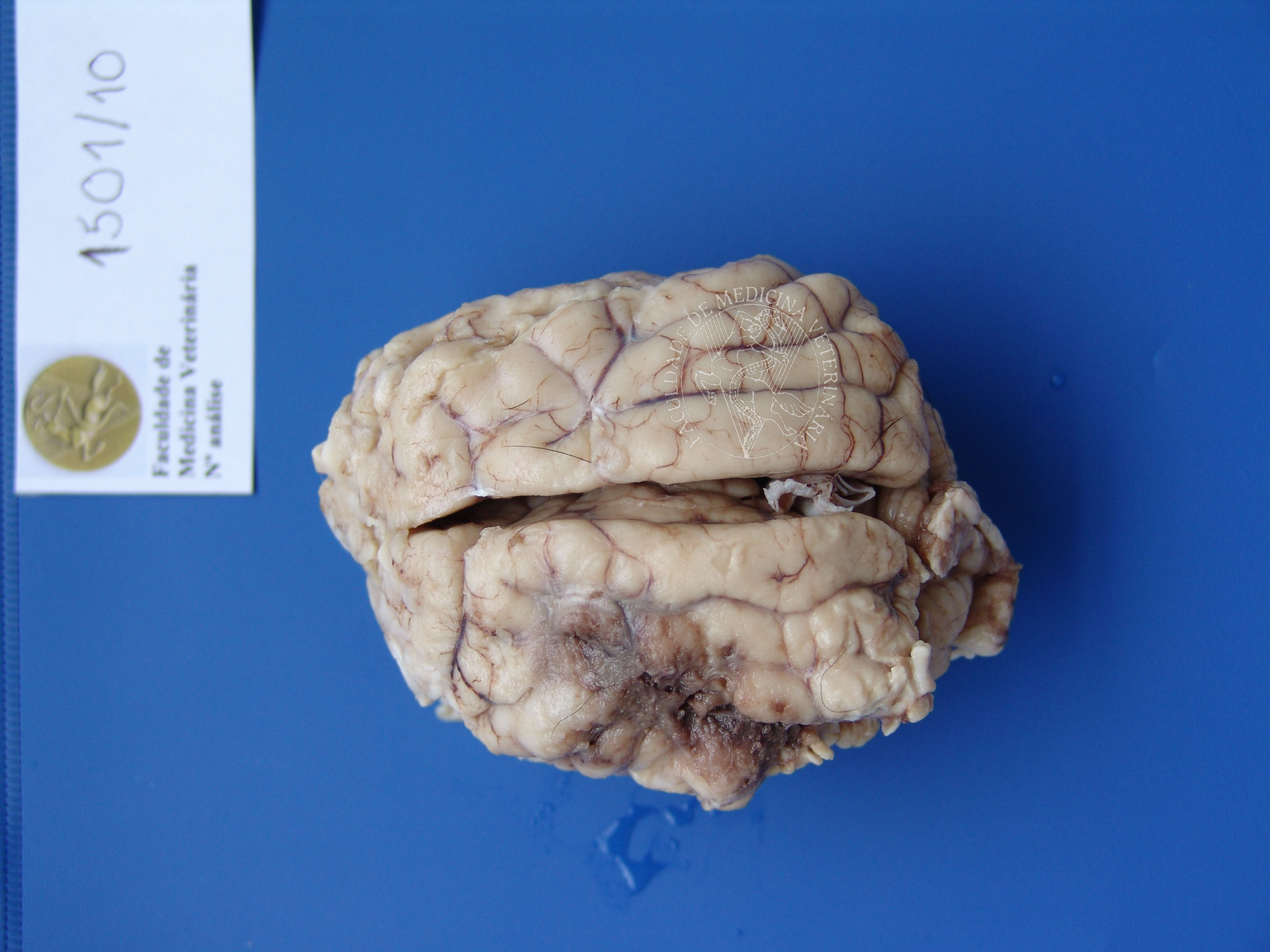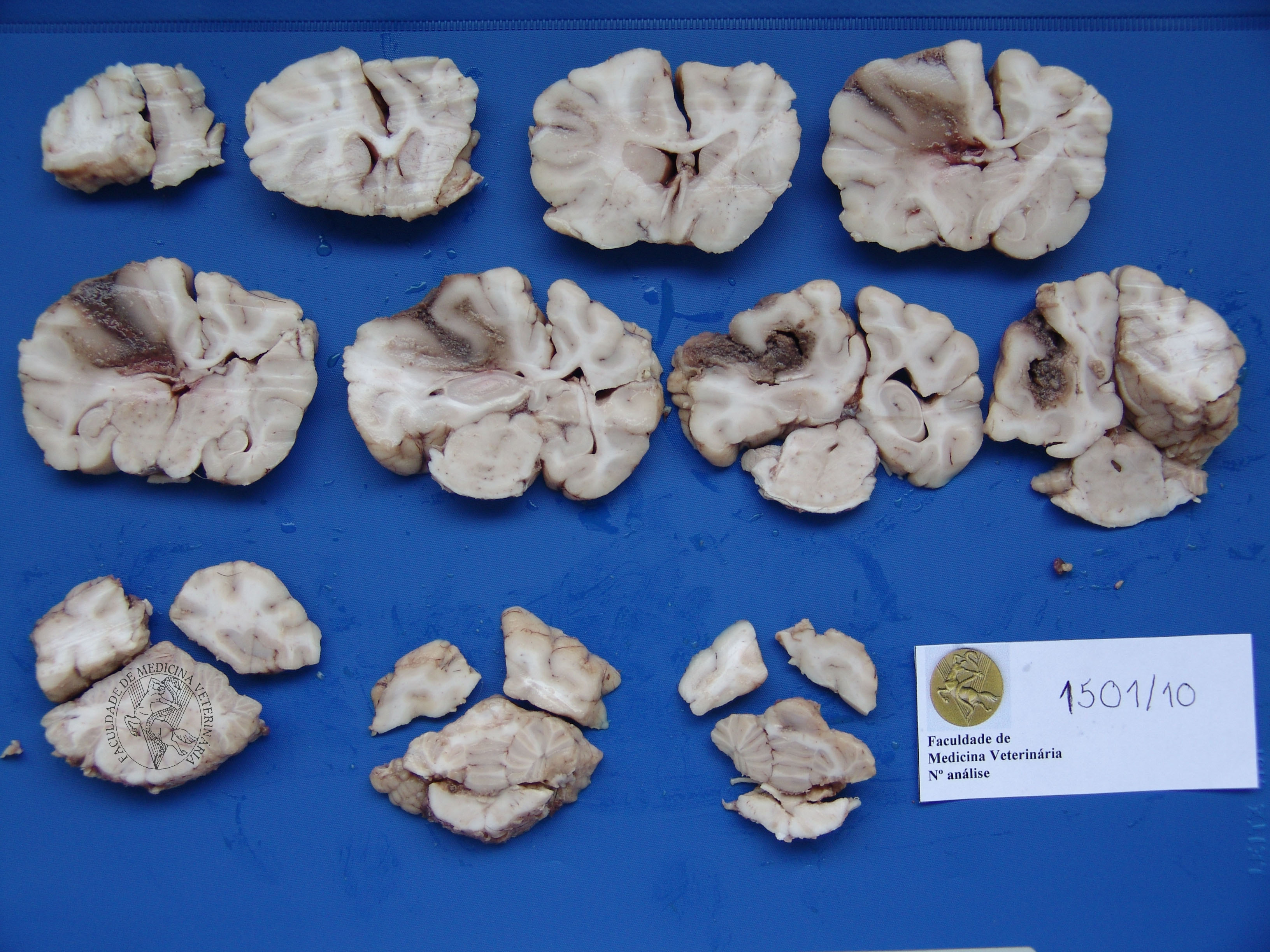

High grade fibrillary astrocytoma in a 7-year-old Fila de São Miguel. The encephalon, shown here after fixation in 10% buffered formalin, exhibits an haemorrhagic mass, about 4 to 5cm in length, affecting both left temporal and parietal lobes, with vaguely perceptible boundaries on serial cuts. Histologically, the mass corresponded to a neoplastic proliferation of round to spindle-shaped astrocytes, sometimes arranged in pseudo-palisade and separated by fibrillary stroma.
 |
 |
 |
Portuguese
English
 |
 |
 |
Loading
Copyright © 2001 by MC Peleteiro. M Pinho & JS Orvalho
design by R Noiva