
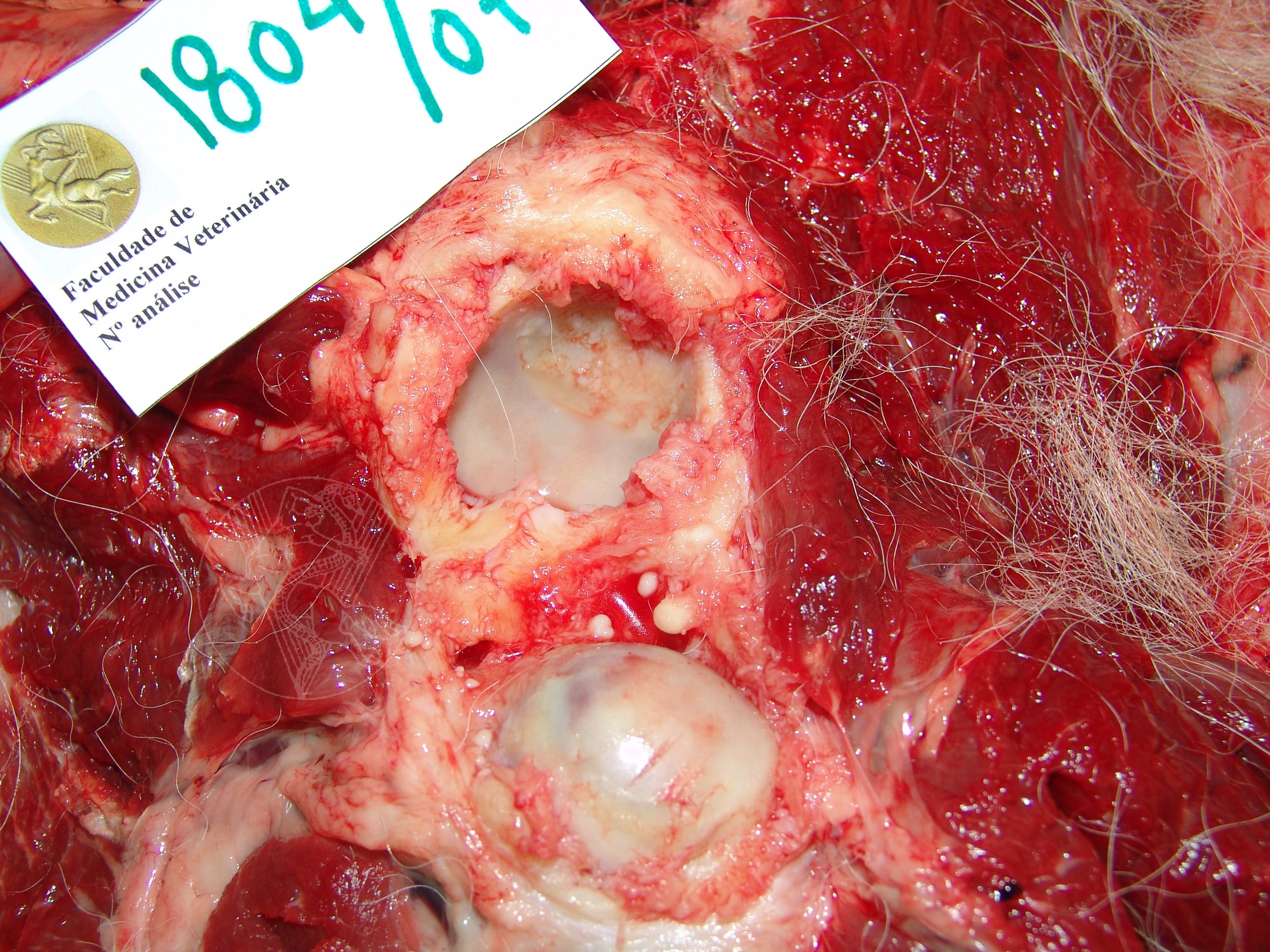
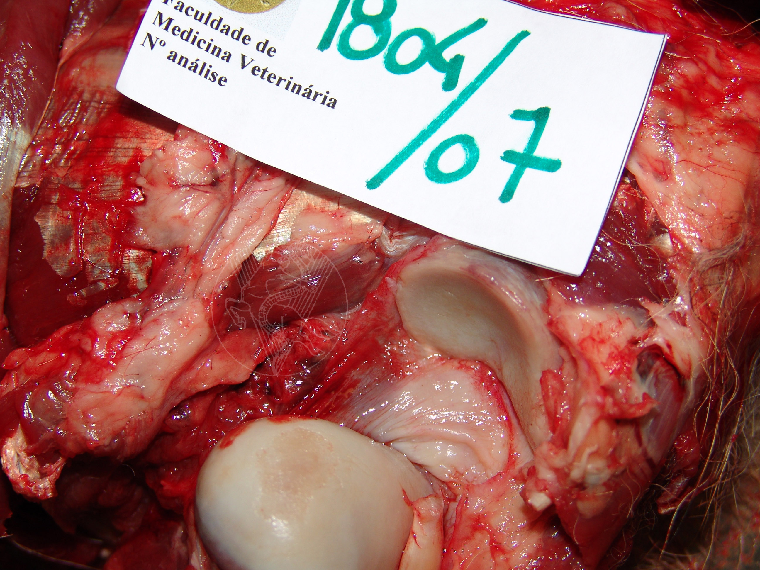
Hip dysplasia in a 9-year-old German Shepherd. Note the marked fibrous thickening of the joint capsule that tries to compensate for the instability generated by the dysplasia. The border where the capsule is fixated on the femur appears nodular and highly irregular. The joint’s surface is heterogeneous, with dark areas around the atrophied hyaline cartilage that enable visualization of the underlying bone tissue. On the right, eburnation (ossification) of the scapular humeral cartilage can be seen.
 |
 |
 |
Portuguese
English
 |
 |
 |
Loading
Copyright © 2001 by MC Peleteiro. M Pinho & JS Orvalho
design by R Noiva