

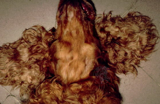
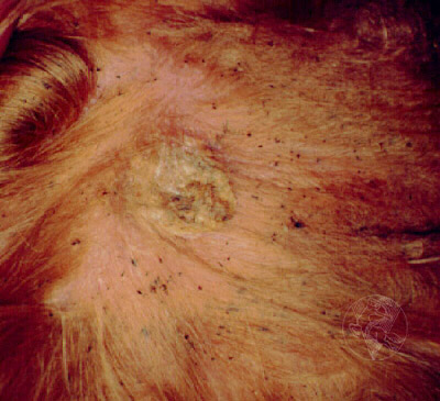
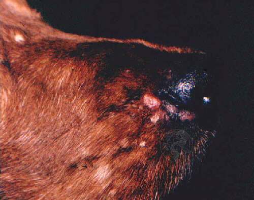
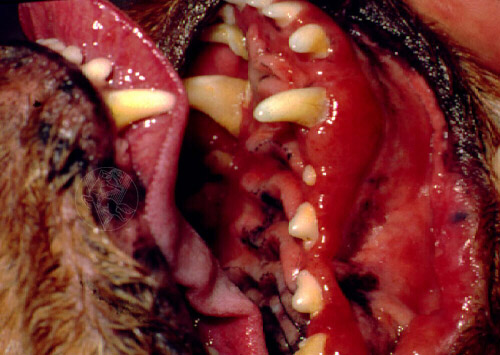
Mandibular lymph node hypertrophy in a case of T-cell cutaneous lymphoma (mycosis fungoides). The image on the right shows a cutaneous plaque corresponding to infiltration by the tumour cells. The fine black dots are flea faeces. Below, on the left, are several small, alopecic cutaneous plaques near the nose. Below, on the right, an image illustrating gingival congestion and hypertrophy is displayed.
 |
 |
 |
 |
 |
Portuguese
English
 |
 |
 |
Loading
Copyright © 2001 by MC Peleteiro. M Pinho & JS Orvalho
design by R Noiva