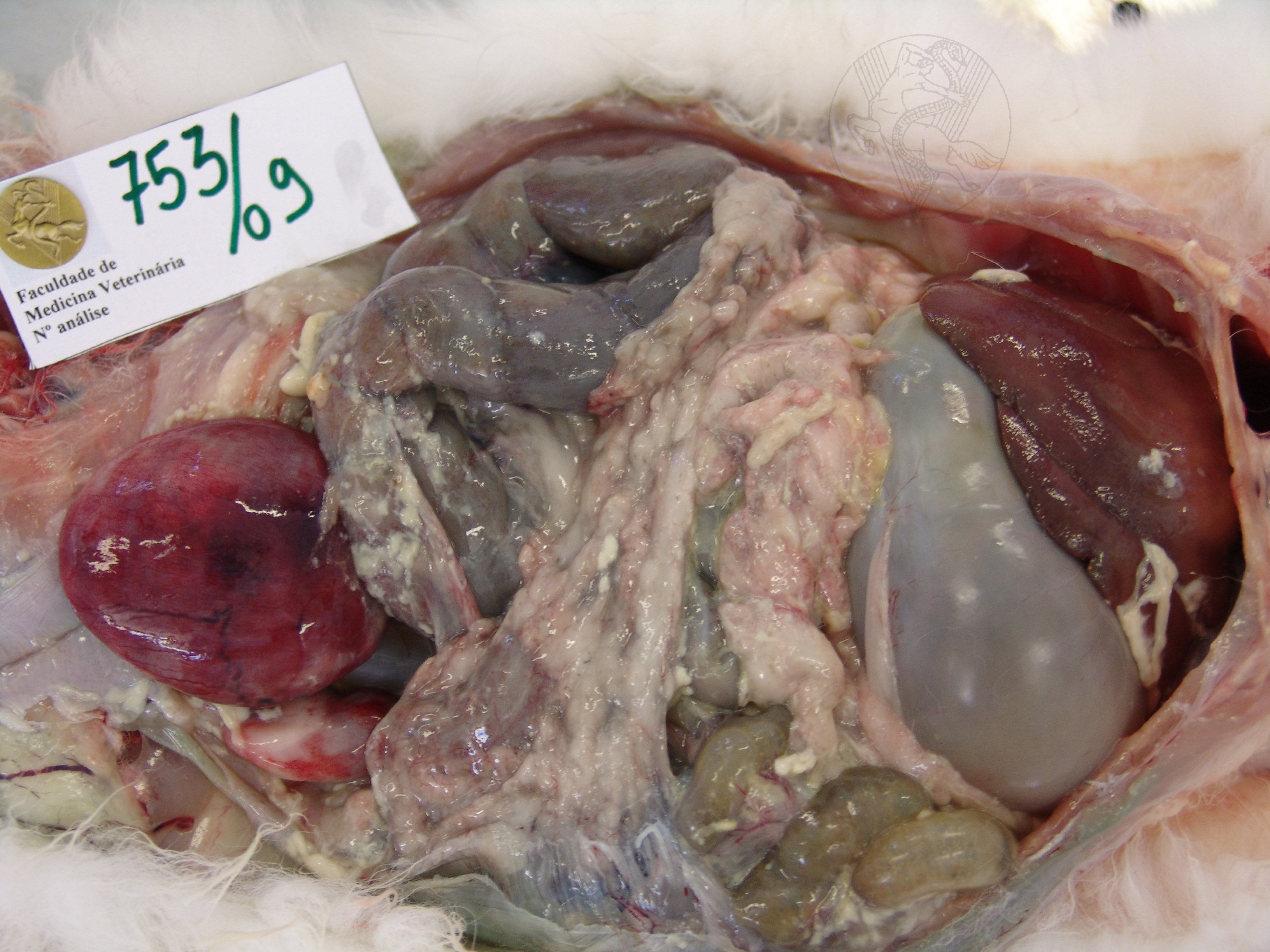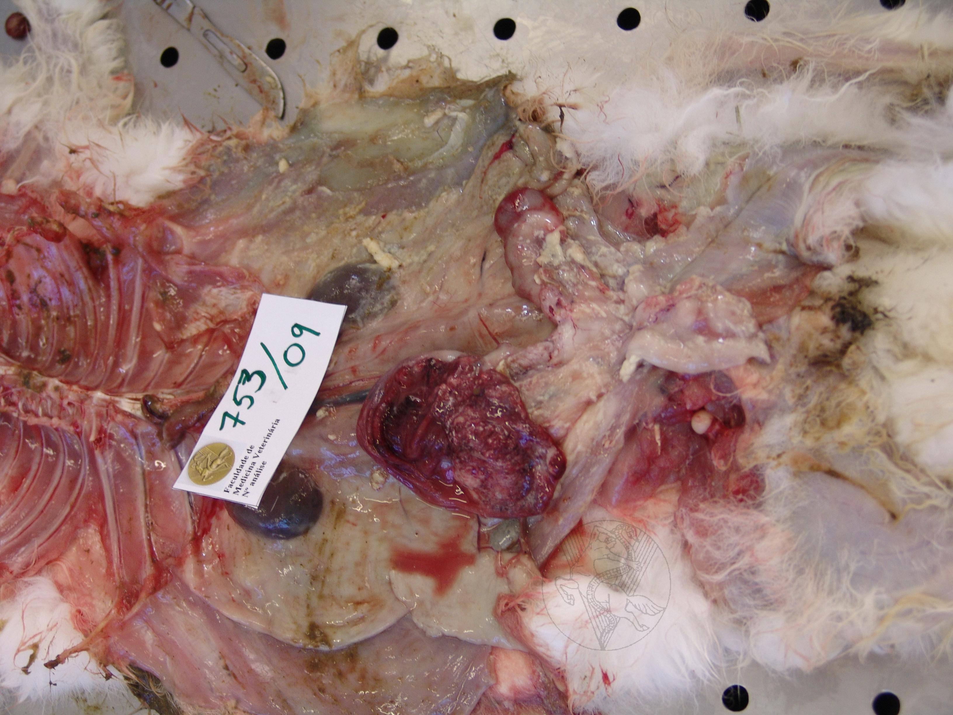

Fibrinous peritonitis secondary to uterine
rupture (not visible in the image) in an 8-year-old doe. On the right-side
image the right uterine horn has been cut open, showing the haemorrhagic
appearance of the uterine wall. A uterine adenocarcinoma was identified
upon histopathological analysis.


