

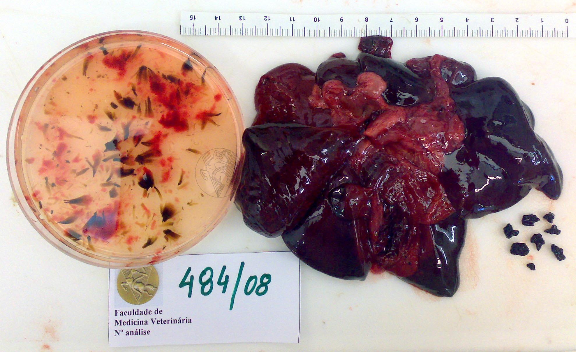
Biliary calculi in a dog. The gallbladder, here completely open, exhibited lesions of chronic hyperplastic cholecystitis, with the lining completely covered with spiculae of solidified mucous identical to the ones seen in the Petri dish, on the left. The gall bladder also contained dark, small, angular calculi that have been placed to the right of the liver.
 |
 |
 |
 |
 |
 |
 |
 |
 |
 |
Portuguese
English
 |
 |
 |
Loading
Copyright © 2001 by MC Peleteiro. M Pinho & JS Orvalho
design by R Noiva