
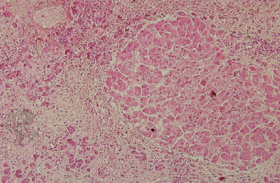
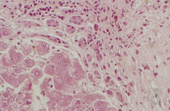
Postdegenerative hepatic cirrhosis. Note the interstitial fibrous tissue hyperplasia and the changes in the structure of the hepatic trabeculae that form a pseudolobule. Within the fibrous tissue, biliary canal hyperplasia can be identified (H&E, 100x). The image on the right shows the hyperplastic perilobular biliary ducts. Note how the peripheral hepatocytes have suffered metaplasia, transforming into lining cells. Although morphologically similar to normal ducts, the new biliary ducts are not functional (H&E, 400x).
 |
 |
 |
 |
 |
 |
 |
 |
 |
 |
Portuguese
English
 |
 |
 |
Loading
Copyright © 2001 by MC Peleteiro. M Pinho & JS Orvalho
design by R Noiva