

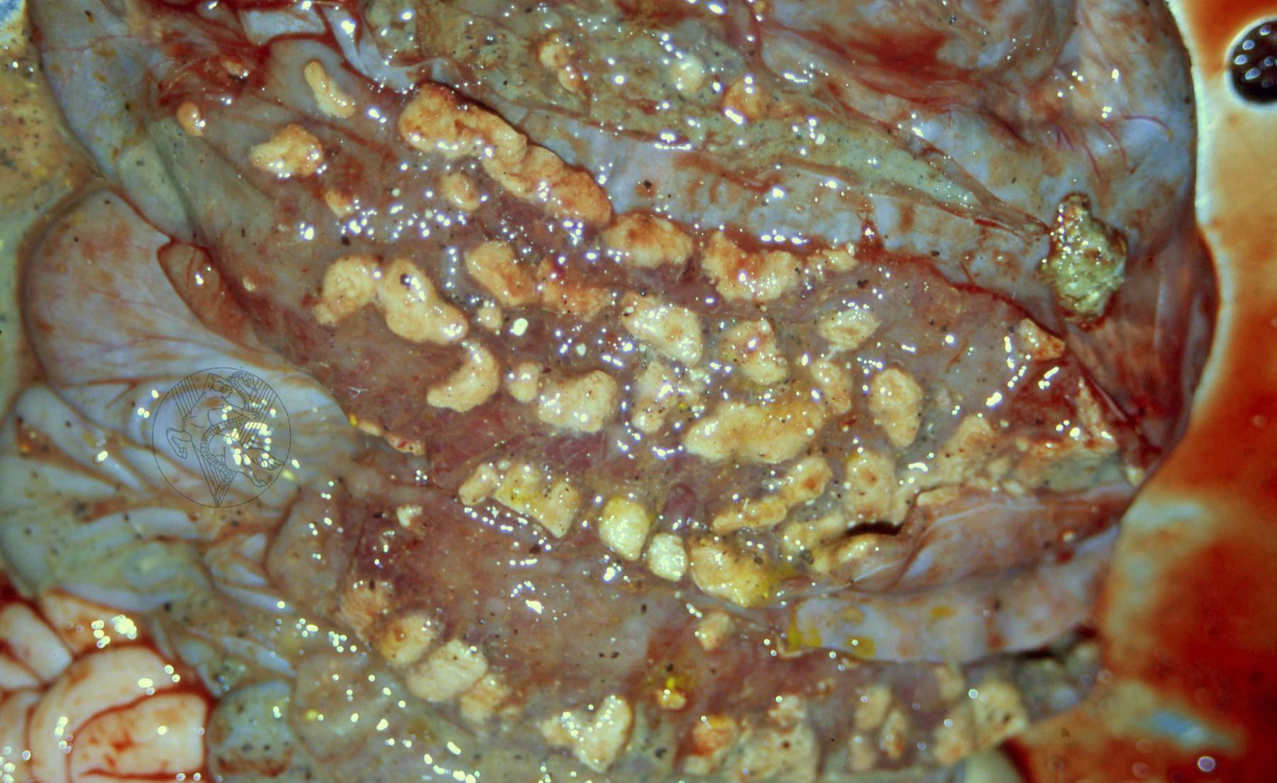
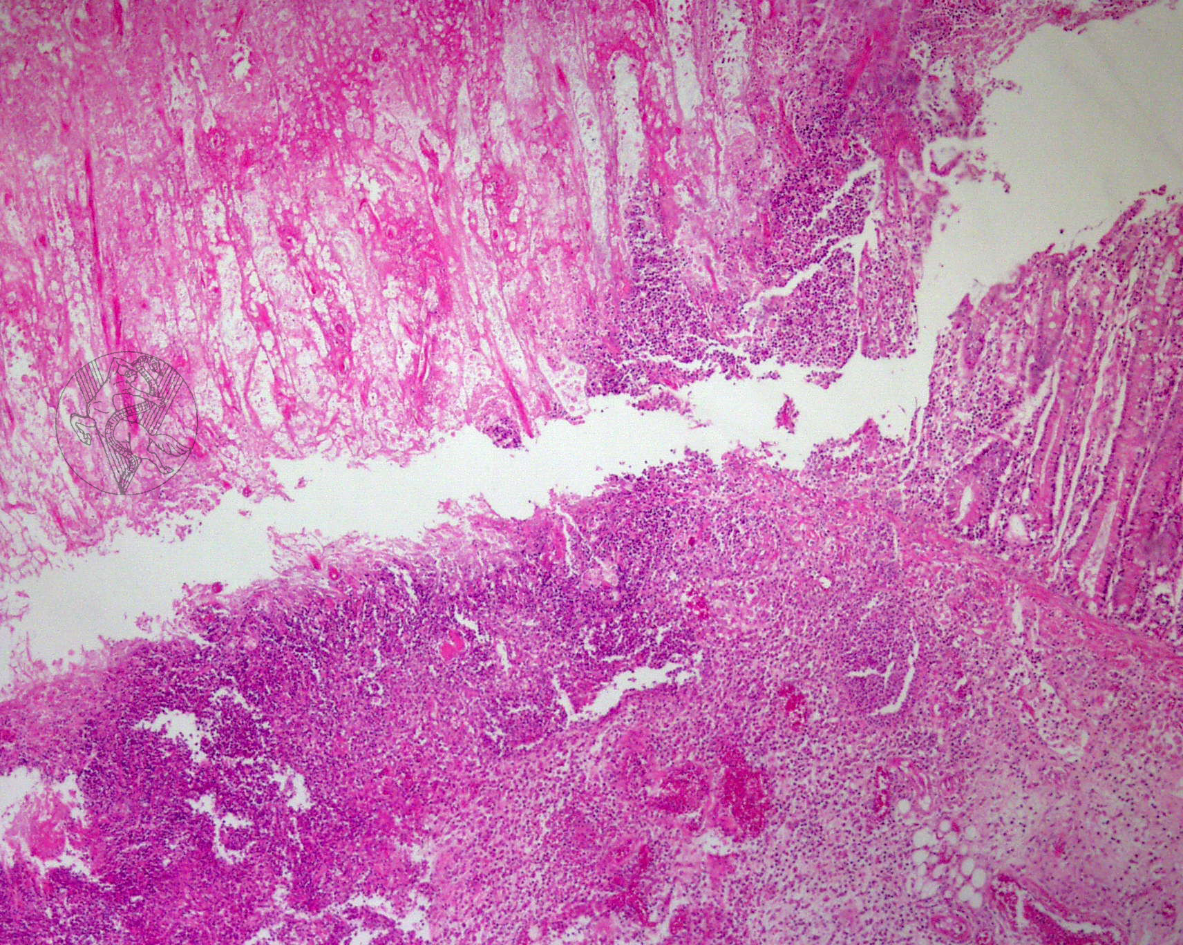
Diphteroid/necrotic enteritis in a pig. Strongly adhered yellowish plaques corresponding to focal fibrinous/necrotic inflammation can be identified on the mucosa. The mucosa has been destroyed and the exudate is strongly attached to the submucosa. On the right, a microscopy image of the lesions can be seen. Note how the necrotic portion of the mucosa (left side of the image) has become detached due to differences in resistance to traction. The intestinal glands (right side of the image) where the mucosa has remained attached do not show any necrosis. There is severe submucosal infiltration.
 |
 |
 |
 |
 |
 |
Portuguese
English
 |
 |
 |
Loading
Copyright © 2001 by MC Peleteiro. M Pinho & JS Orvalho
design by R Noiva